+2,100 students
Learning with VRDI simulators across Latin America and Spain

+11 years
Developing with virtual reality technology

+580,000 assessments
Completed using VRDI simulators.
Section: Bring theory into practice—safely and realistically
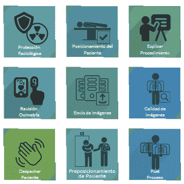
DoReality VRDI replicates the end-to-end patient-care workflow for MRI, CT, and X-ray, strengthening hands-on learning.
- Full clinical cases: From patient intake to image interpretation.
-Multiplatform: Compatible with PC and Virtual Reality.
-Unlimited practice: Repeat procedures with zero risk to patients.
-Instant feedback: Builds technical skills and communication.
-Regular updates and upgrades: Ongoing content and feature improvements.
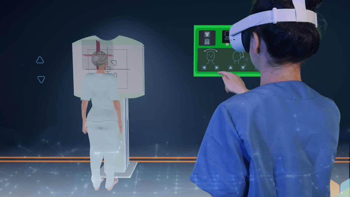

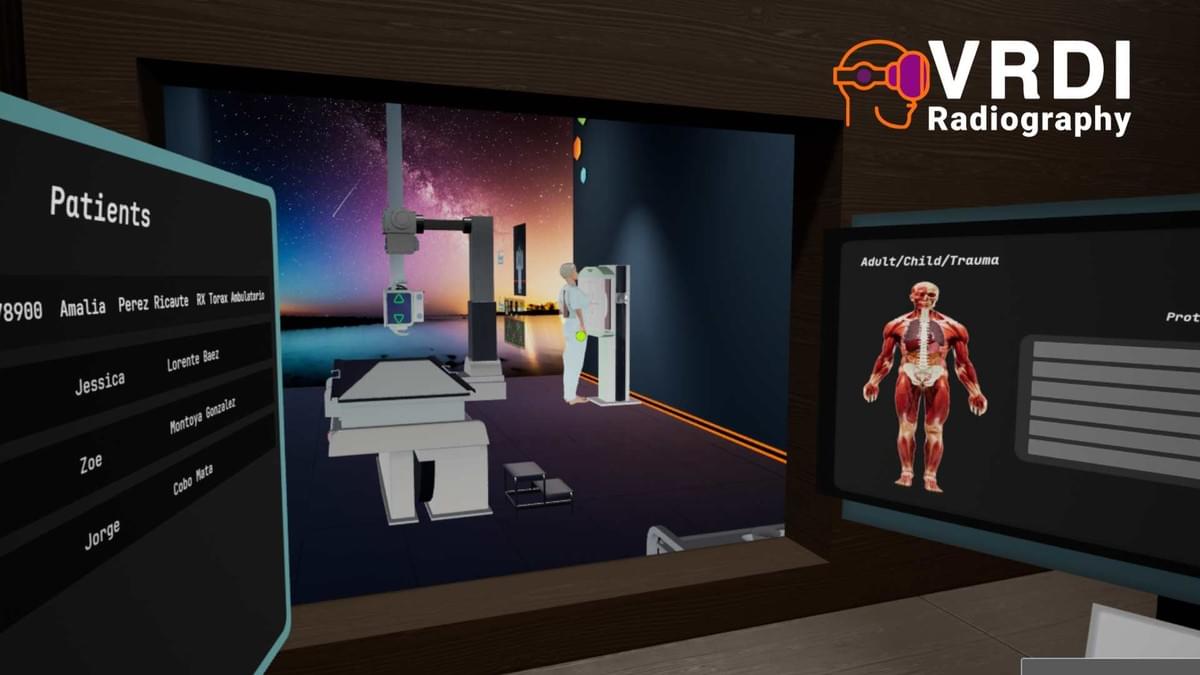
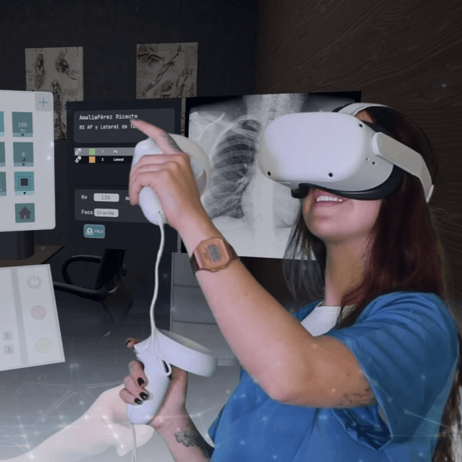
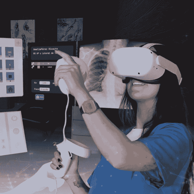
X-Ray Simulator
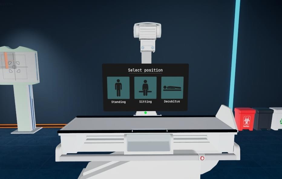
Training in conventional radiology: patient positioning, parameter selection, and image acquisition.

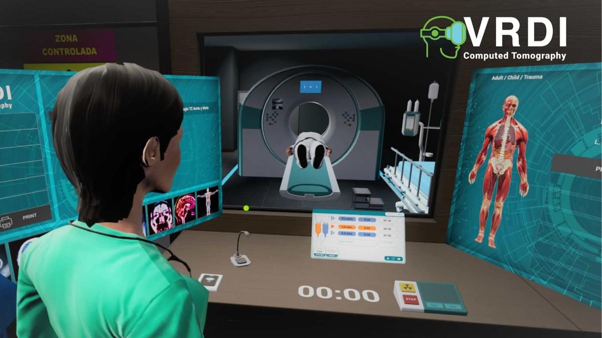
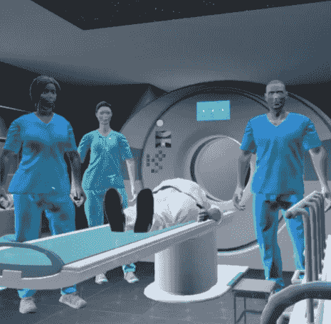
CT Simulation
End-to-end procedural simulation—from patient intake to viewing cross-sectional images.

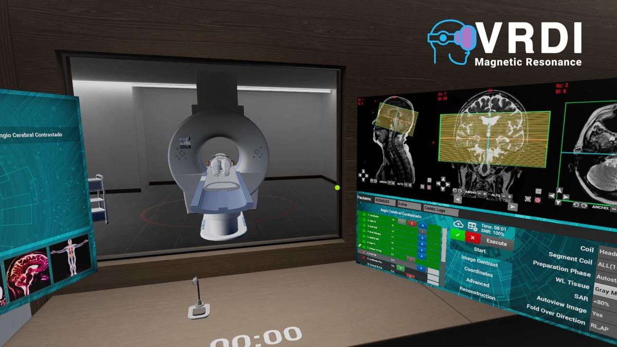
MRI Simulator
Guided and self-directed training across technical workflows, patient interaction, and results analysis.
What do our simulators offer?
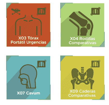
Cases
Clinical cases at varying levels of complexity to strengthen hands-on learning and critical analysis.
Situations
Decision-making scenarios that develop clinical judgment and problem-solving in a controlled environment.
Characters
Virtual patients and healthcare staff that recreate real-world interactions, fostering both technical and soft skills.
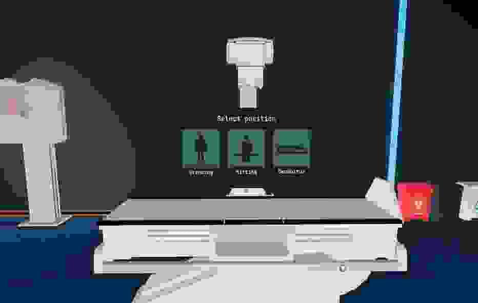
Environments
Facilities, equipment, and virtual software interfaces that faithfully reproduce the clinical setting for safe, immersive practice.
Indicators
End-to-end performance traceability to track progress, identify strengths, and support formative assessment.
During the simulation
All learner decisions are captured and analyzed—enabling adaptive learning paths, full traceability, and evidence-based feedback.
These interactions also allow faculty and institutions to monitor progress, evaluate simulator usage, and assess mastery of the clinical workflows involved.Recomendación de copy (directo y corporate): usa end-to-end diagnostic workflow, evidence-based feedback y la primera mención expandida de RIS (Radiology Information System) para elevar claridad y credibilidad.
Expected Learning Outcomes
Competencies students will develop in the simulation

Understand the end-to-end diagnostic workflow: from administrative intake through case closure in RIS-type systems.

Operate confidently in virtual clinical environments: identify the layout, functions, and dynamics of the exam room and the control room.

Operate simulated biomedical equipment: use software interfaces correctly.

Apply clinical protocols: tailor parameters and technical decisions to each case.

Interact with virtual patients and clinical staff: develop communication and clinical-care skills

Understand image post-processing workflows, among other learning objectives.

Reinforce theory through practice: make decisions, learn from safe-to-fail errors, and repeat procedures to consolidate learning.
What they say about our simulators
Educators using VRDI in class share their experience.

“A highly detailed instructional tool. It’s 100% interactive, lets you increase the difficulty level, and is quickly adopted by users.”
— Emilio Garcés - Colombia
Professor of Diagnostic Imaging

“The CT simulator brings students into the full axial-tomography workflow: from patient intake and identification to positioning and interpreting real pathological images. With this simulator, students engage in the entire procedure through guided gameplay. It’s a very useful assessment tool that verifies learning.”
— María Isabel Rojo - Spain
Professor of Diagnostic Imaging and Nuclear Medicine